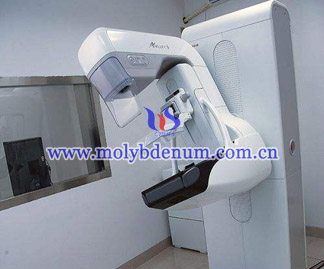
Common Breast X-ray Findings

The performances of common breast cancer in the X-ray molybdenum sputtering target are as follow:
1. Dust carcinoma in situ: this type account for 20%~30% in the census, calcification is the main reason that 90%DCIS was found.
Pathology: the DCIS is derived from small dust breast cancer and the cancer cell was confined with the duct un-invasion the base level.
Types: acne and non-acne type
Clinical manifestation: touch painless mass, partly with Paget's disease
Typical X-ray mammography:
1)The mass ca show a “v” shape distribution with calcification
2) In first quadrants the roundness and irregular cluster sample distribute mass calcification or distribute in lots of small cluster sample with calcification.
2. Invasive dutal carcinoma: this breast cancer is most common which account for 60% of breast cancer.
Pathology: the cancer cell breaks through basement membrane and continued to infiltrate interstitial tissue.
Clinical manifestation: infiltrative growth, obscure boundary, no encapsulated, rarely see hemorrhage and necrosis.
Typical X-ray mammography:
1) pure mass
2) simple calcification
3) structure distortion
4) mass with calcification
5) negative mammograms
3. Invasive lobular carcinoma: this cancer rank second in second primary breast cancer which account for 8%--14%, middle malignant. And characteristics of it are mainly multifocal, multicentricity and ambi-growing.
Pathology: cancer cell has small size, morphology more consistent, less cytoplasm. And it often shows single cord and can be liner. The cancer cell around tube or lobular shows concentrically structure and molybdenum-like structure.
4.Mucinous adenocarcinama: The mucinous adnocarcinama usually appears in postmenopausal and older women. And the tumor grow slow and metastasis night.
Pathology: large tumor, border clearance, irregular shape, section showing gelatinous, under the microscope interstitial has rich mucus and cancer cell separated into volmer-weber. There is vacuole in cytoplasm and nuclear small and round, less split, engrain, often biased towards one side.
Typical X-ray mammography:
1) uncalcification mass is most common, other features are: large tumors, clear state, located in the edge of the gland, or showing small lobulated, few edge blur, infiltration, high density.
2) rare calcification
3) limitations opacities
5. Medullary carcinoma: this cancer common in young people and it has large tumor usually in 4-6cm. Besides, it often exists in deep central of breast showing spherical right nodular, border clearance, soft, usually common bleeding and necrsis.
Pathology: cancer cell has large size, different form, showing expansive growth, abundant cytoplasm, large vacuolization nucleus, mitotic more common, less lymph node matastasis.
Typical X-ray mammography:
1) the round mass without calcification often showing in the deep breast
2) the mass showing small lobule, infiltrating the edge
3) the mass showing equal gland density


